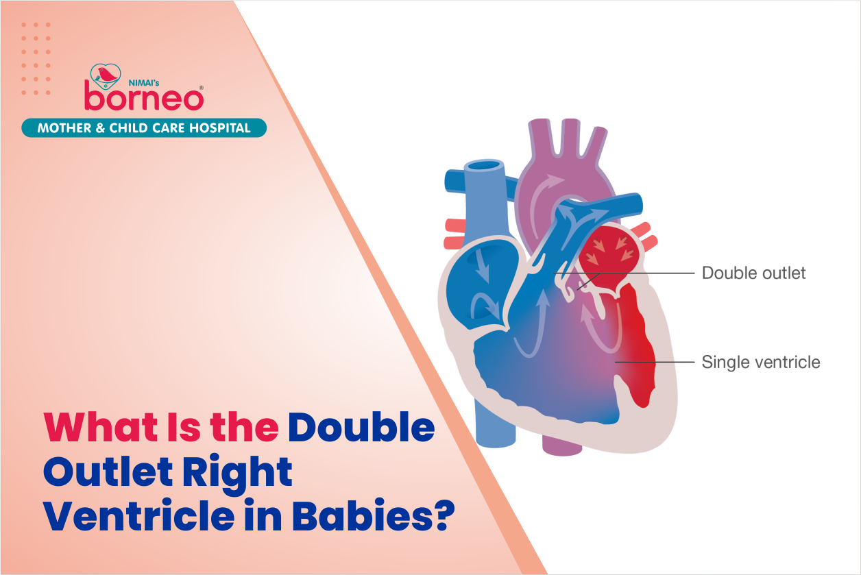
What Is The Double Outlet Right Ventricle And What Does It Look Like
A double outlet right ventricle is a heart defect that occurs when there are two separate, large openings in the wall between the right ventricle and the pulmonary artery. This heart defect can cause a number of different problems, but it’s most often associated with an infant who has been born prematurely. . Some of the complications include chronic lung disease, recurrent infections, and even death. This type of heart defect is typically detected as an infant ages due to the following:
Shortness of breath during a routine physical examination
Difficulty feeding or swallowing
Poor growth and weight gain
A pulse that’s difficult to detect on a regular basis.
The Importance of a Double Outlet Right Ventricle in Infants and When Does it Become a Problem
Dorsoventral thoracic deformity leaves the heart at the top of the chest and it is shifted to the left and down in a downward position. This can result in an abnormal placement of the heart, leading to a condition called extreme right ventricular hypertrophy. The resulting enlarged right ventricle pumps blood through an opening (hole) in the normal wall between it and the left ventricle, creating a very high pressure to push blood through this hole. This increased pressure can cause great strain on major veins near where these veins enter into your lungs, as well as other problems related to the lungs and heart. This deformity can be differentiated from other congenital heart defects such as hypoplastic left heart syndrome, the ventricular septal defect, or a pulmonary atresia.
The 4 Stages of Development to Understand the Difference Between Normal & Abnormal Development and Why It Matters
The right ventricle system is an important part of the heart and helps to pump oxygenated blood to the rest of the body. A double outlet right ventricle is a condition where there are two right ventricles instead of one and this can be potentially dangerous.
It is important to know when a dorsoventral thoracic dextroposition (DVT) is present in infants because it can lead to serious complications such as cardiogenic shock, pulmonary edema, and death.
A dorsoventral thoracic dextroposition (DVT) happens when a child has their chest tilted towards their back instead of towards their belly button. This can happen in normal development or if there are other congenital conditions that cause this to happen.
What Tests Are Involved in Treating the Double Outlet Right Ventricle?
The right ventricle is a large air sac that is located in the right side of the heart. It contains two blood vessels, the pulmonary artery and the azygos vein. The right ventricle pumps blood to the lungs through these two blood vessels.
● The most common test used in diagnosing right ventricular outflow tract obstruction is echocardiography. This test uses sound waves to create an image of your heart and its chambers. It can also be used to measure how well your lungs are working, which can help determine whether you have congestive heart failure or not. Right ventricular outflow tract obstruction refers to a narrowing or closure of one or both of the arteries that branch off from the main pulmonary artery that go into your lungs. If a person has right ventricular outflow tract obstruction, this means that the heart’s workings are not as efficient in sending oxygenated blood to the lungs.
How to Diagnose the Child with a Double Outlet Right Ventricle and When to Expect Surgery or Treatment Options
A diagnosis of a double outlet right ventricle indicates that there is an increased chance for a child to develop life-threatening complications, such as heart failure. The most common treatment option for this condition is surgery to close one of the outlets. A child with a double outlet right ventricle can also be treated with medications and supportive care, which are less invasive than surgery. The most common finding on a chest X-ray is the size of the heart. Abnormally large hearts make it difficult for children to get enough oxygen into their blood.
A diagnosis of a large heart can be an indication of either congestive heart failure or cardiomyopathy, which is where the pumping ability of the heart is decreased due to structural changes in one or more chambers. Chest X-rays are not diagnostic, so they are often repeated over time and during different phases of development in order to determine if a larger size leads to other abnormalities in development. An enlarged heart may directly cause symptoms, such as shortness of breath. Abnormally large hearts may lead to congestive heart failure, which can include shortness of breath during activity and fluid build up in the lungs.
Children with a large heart are also at an increased risk for atrial septal defect (ASD), where there is a hole in the wall separating two chambers in the heart. A child with a large chest X-ray may have pulmonary venous hypertension (PVH), which is when blood flow back to the lungs becomes constricted due to pressure from high blood volume and causes backflow.
The most common causes for a heart abnormality in children are congenital heart disease, cardiomyopathy and congenital heart disease.
Congenital abnormalities may be present from birth or may develop over time as the child gets older. If a significant cause is not identified by 18 months of age, the child should be considered to have cardiomyopathy. Cardiomyopathy is where the pumping ability of the heart is decreased due to structural changes in one or more chambers of the heart. An enlarged heart may directly cause symptoms such as shortness of breath and fatigue.In most cases, the children are being seen because of chest pain that is not relieved by rest or medication. A CT scan may be ordered for ruling out other causes for the chest pain. Other tests may include an echocardiogram to evaluate how well the heart is pumping blood and investigate if there are abnormalities in the heart valves or chambers that were seen on a chest X-ray. If no other cause is found, a cardiology evaluation will be planned to assess whether further testing may include cardiac catheterization which can diagnose additional defects in any or all of the heart’s chambers.


